

Behind every symptom, whether a swollen leg or an unexplained ache in the abdomen, there is a question about what is happening inside the body. An ultrasound test helps answer it by revealing the shape, size and position of organs and tissues in real time.
The Colour Doppler test shows how blood moves through the vessels supporting those structures, including its direction and speed. When a doctor requests one or both, the aim is to match the scan to the clinical question, so that findings are precise and the next step in care is clear.
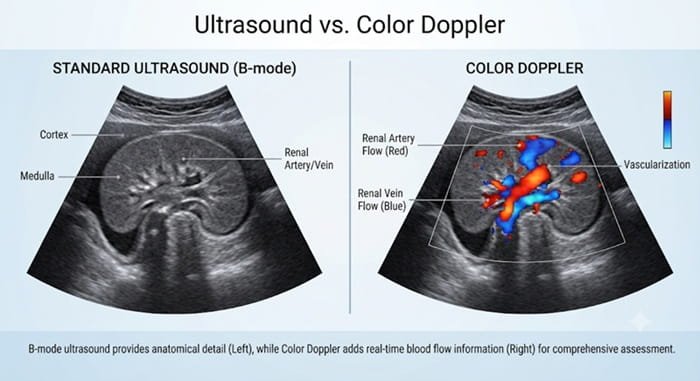
This guide explains how the two tests differ, their common uses and when each is recommended.
Ultrasound is an imaging test that uses high-frequency sound waves to create real-time images of internal organs and tissues. It is commonly used to examine the abdomen, pelvis, thyroid, kidneys, liver, gallbladder, muscles and soft tissues as well as to monitor pregnancy.
An ultrasound test can identify cysts, stones, fluid collections, organ enlargement and other structural abnormalities. Because it does not use ionising radiation, it is suitable for patients of all age groups, including unborn babies, children and older adults.
Colour Doppler is an advanced ultrasound technique that evaluates moving blood cells. It uses colour mapping to display the direction and relative speed of blood flow through arteries and veins.
The colours do not directly indicate whether a finding is favourable or unfavourable. They assist the radiologist in interpreting flow patterns and determining whether circulation appears normal.
Colour Doppler is often combined with a standard ultrasound in a single examination, sometimes called a duplex ultrasound. This allows the radiologist to assess the structure of a vessel and the blood moving through it at the same time.
The following table summarises the principal differences between the two tests:
| Feature | Standard Ultrasound | Colour Doppler |
| Main purpose | Creates images of organs, tissues and structures | Assesses blood flow within vessels and organs |
| What it shows | Size, shape, position, cysts, stones, masses, fluid | Direction, presence, absence and relative speed of blood flow |
| Common use | Pregnancy scans, abdominal pain, thyroid or pelvic evaluation | Suspected clots, narrowed arteries, varicose veins, organ blood supply |
| Typical output | Grey-scale anatomical images | Colour-coded flow images, often with grey-scale images |
| When recommended | When a structural problem is suspected | When circulation, vessel blockage or abnormal flow is a concern |
Doctors commonly recommend a standard ultrasound test in the following situations:
Colour Doppler is commonly recommended in the following clinical situations:
Both tests follow a similar and simple process. Before booking an appointment, patients searching on the web for ‘ultrasound scan near me’ should check the availability of the required scan and confirm preparation instructions.
Here is what you should be prepared for:
A standard ultrasound test shows what is inside the body, while colour Doppler shows how blood is moving through it. Neither replaces the other, and each becomes more useful when the question being asked is clear. Your doctor will recommend the right scan based on your symptoms and medical history, and in many cases both are done in a single visit. Because neither involves radiation, they are among the safest ways to look inside the body.
Diagnostic centres such as Vijaya Diagnostics offer both ultrasound and Doppler imaging, so patients can get timely answers under proper medical guidance. If you have been advised to undergo either of the tests, speak with your doctor about what to expect and what the results mean for you.
References